Subcutaneous muscle neck (platysma) tightens the skin of the neck and part of the sternum, and also shifts the corner of the mouth forward and down. The muscle is a thin wide plate located under the skin of the neck and partially under the skin of the face. The point of its beginning is in the subclavian region at the fascia of the large chest and deltoid muscle, and the attachment point is the edge of the lower jaw, chewing fascia and the corner of the mouth.
The sternocleidomastoid muscle (m. sternocleidomastoideus) (Fig. 90, 95, 96) with bilateral contraction throws its head back, and with one-sided it tilts its head to its side (to the side on which the muscle contracts) and turns it in the opposite direction.
The muscle is a thick long strand with two heads, going obliquely from the mastoid process through the neck to the sternoclavicular joint. The lateral head of the muscle has the anterior surface of the sternum handle as its starting point, and the medial head has the sternal end of the clavicle. The muscle is attached to the mastoid process and the lateral section of the upper nuchal line.
 | Rice. 96. Superficial, median and deep muscles of the neck (side view): 1 - stylohyoid muscle; |
 | Rice. 97. Median and deep muscles of the neck (side view): 1 - maxillofacial muscle; |
The phrase "head off your shoulders" from the point of view of anatomy is not entirely correct, since between the head and shoulder girdle such a part of the body as the neck is located. The basis of the neck is made up of seven vertebrae interconnected. The neck is a very mobile part of our body, which is ensured by the special structure of the vertebrae and the presence of muscles.
All neck muscles are divided into three groups: superficial, median and deep muscles.
Superficial muscles include muscles such as:
- The subcutaneous muscle of the neck, starting in the region of the second rib and ending at the edge of the lower jaw. Contracting, it stretches the skin of the neck and chest, and also lowers the lower jaw, pulls the corner of the mouth outwards and downwards.
- The sternocleidomastoid muscle begins with its lateral head from the sternal end of the clavicle, and medially from the anterior surface of the sternum handle, and ends on the mastoid process of the temporal bone. Contracting on one side, it tilts its head to its side and turns its faces to the opposite side, and with a bilateral contraction, it throws its head back and pushes it forward a little.
The median muscles of the neck muscles include:
Suprahyoid muscles:
- Digastric muscle - starts with two heads from the digastric fossa of the lower jaw and is attached to the temporal bone. Contracting, lowers the lower jaw, pulls the hyoid bone.
- The stylohyoid muscle originates from the styloid process of the temporal bone and ends on the body and greater horn of the hyoid bone. Contracting, pulls the hyoid bone back.
- The maxillohyoid muscle begins on the lower jaw and ends on the body of the hyoid bone. Contracting, it shifts the hyoid bone up, back and outward.
- The geniohyoid muscle begins on the mental spine of the lower jaw and ends on the anterior surface of the body of the hyoid bone. Contracting, it pulls the hyoid bone forward and upward and participates in lowering the lower jaw.
Hyoid muscles:
- The sternohyoid muscle begins on the posterior surface of the clavicle, the articular capsule of the sternoclavicular joint and the manubrium of the sternum, and ends on the body of the hyoid bone. Contracting, lowers the hyoid bone down.
- The sternothyroid muscle begins on the surface of the 1st rib, the handle of the sternum, and ends on the larynx, attaching to the oblique line of the thyroid cartilage. Contracting, lowers the larynx down.
- The thyroid-hyoid muscle begins along with the previous one and ends on the large horn of the hyoid bone. Contracting, brings together the hyoid bone and the larynx.
- The levator thyroid muscle arises from the thyroid-hyoid muscle and ends at the capsule of the thyroid gland. The function is visible from the name.
- The scapular-hyoid muscle begins with the upper abdomen on the body of the hyoid bone, and the lower one fuses with the fascial sheath of the neurovascular bundle of the neck.
The deep muscles of the neck are divided into lateral and prevertebral muscle groups.
The prevertebral muscle group includes:
- long muscle head, starting at the anterior tubercles of the III-VI cervical vertebrae and ending on the basilar surface of the occipital bone. Contracting, the longus capitis muscle tilts the head and neck forward.
- The longus muscle of the neck is divided into three parts:
- A) medially-vertical, starting from the bodies of the fifth cervical - third thoracic and ending on the bodies of the second or third cervical vertebrae and the atlas.
- B) the upper oblique part, starting from the costal-transverse processes of the II-V cervical vertebrae and ending on the body of the second cervical vertebra.
- C) the lower oblique part, starting from the upper three thoracic vertebrae and ending on the costal-transverse processes of the last three vertebrae.
- Contracting, the long muscle of the neck tilts the neck forward and to its side.
- The anterior rectus capitis originates from the transverse process and the lateral mass of the atlas and ends at the base of the occipital bone. Contracting on one side, tilts the head forward, and with two-sided contraction forward.
- The lateral rectus capitis begins at the transverse process of the atlas and ends at the jugular process of the occipital bone. Shrinking, he tilts his head.
The lateral group of deep neck muscles include:
- Anterior scalene muscle - begins the third or fourth cervical vertebrae and ends at the first rib. Contracting on one side, pulls the neck to its side, with bilateral contraction, the neck forward.
- The middle scalene muscle starts from the six upper cervical vertebrae and ends on the surface of the first rib. Contracting, tilts the neck forward.
- The posterior scalene muscle starts from the fifth - fourth cervical vertebrae and ends on the surface of the second rib. Contracting, tilts the cervical spine forward.
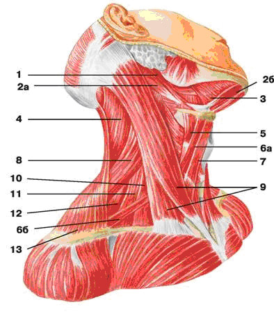
The muscles of the neck are topographically divided into superficial, median and deep groups. median group subdivided into muscles located above and below the hyoid bone. In the group of deep muscles, lateral and medial (prevertebral) groups are distinguished.
Superficial muscles
1. Subcutaneous muscle The neck is located as a thin wide plate under the skin of the neck and part of the face. Beginning: in the subclavian region from the fascia of the deltoid and large chest muscle; attachment: corner of the mouth, edge of the lower jaw, masticatory fascia.
Function: lifts the skin of the neck, partly the chest, pulls the corner of the mouth outwards and downwards.
2. Sternocleidomastoid muscle forms a long thick cord, obliquely crossing the neck from the mastoid process to the sternoclavicular joint. Has two heads. Beginning: medial head - anterior surface of the sternum handle, lateral - sternal end of the clavicle; attachment: mastoid process and lateral section of the upper nuchal line.
Function: with unilateral contraction, it turns its head in the opposite direction, tilts it to its side, with bilateral contraction, it throws its head back.
Superficial muscles of the head and neck
Middle group. Suprahyoid muscles
1. Digastric has two bellies - anterior and posterior, connected by a tendon bridge. Beginning: anterior abdomen - digastric fossa of the lower jaw, posterior - mastoid notch of the temporal bone; attachment: both abdomens pass into a tendon, which is attached to the body of the hyoid bone.
Function: lowers the lower jaw, pulls it back. With a fixed lower jaw, it raises the hyoid bone.
2. Stylohyoid muscle- thin fusiform muscle. Origin: base of the styloid process of the temporal bone; insertion: body and greater horn of the hyoid bone.
Function: pulls the hyoid bone up, back and outward.
3. Maxillofacial muscle flat, connecting with the muscle of the same name on the opposite side, forms the bottom of the oral cavity (diaphragm of the mouth). Beginning: maxillary-hyoid line of the lower jaw; attachment: the posterior bundles are attached to the anterior side of the hyoid bone, the main part meets the fibers of the opposite muscle of the same name, forming the maxillo-hyoid suture of the diaphragm of the mouth.
Function: raises the hyoid bone, when it is fixed, lowers the lower jaw.
4. Geniohyoid muscle located above the jaw-hyoid muscle. Origin: mental spine of the lower jaw; insertion: anterior surface of the body of the hyoid bone.
Function: pulls up and forward the hyoid bone, when it is fixed, lowers the lower jaw.
Infrahyoid muscles
1. Scapulohyoid muscle long, thin, divided by an intermediate tendon into two bellies. Beginning: upper abdomen - lower edge of the hyoid bone, lower - upper edge of the scapula, upper transverse ligament; attachment: both bellies are connected to each other by a tendon bridge.
Function: with a fixed scapula, it pulls the hyoid bone down and outward, and also pulls the sheath of the neurovascular bundle of the neck, thereby expanding the lumen of the internal jugular vein.
2. Sternohyoid muscle. Origin: posterior surface of the clavicle, manubrium of the sternum, capsule of the sternoclavicular joint; attachment: the lower edge of the body of the hyoid bone.
Function: pulls the hyoid bone down.
3. Sternothyroid muscle. Origin: posterior surface of the manubrium of the sternum, cartilage of the 1st rib; attachment: oblique line of the thyroid cartilage of the larynx.
Function: pulls the larynx down.
4. Thyrohyoid muscle. Origin: oblique line of the thyroid cartilage; insertion: body of the hyoid bone.
Function: brings together the hyoid bone and the larynx, with a fixed hyoid bone raises the larynx.

Muscles of the head and neck; side view. 1 - temporalis muscle(m.temporalis); 2 - occipital-frontal muscle (m. occipitofrontalis); 3 - circular muscle of the eye (m. Orbicularis oculi); 4 - large zygomatic muscle (m. zygomaticus major); 5 - muscle that lifts the upper lip (m. Levator labii superioris); 6 - muscle that raises the corner of the mouth (m. Levator anguli oris); 7 - buccal muscle (m. buccinator); 8 - chewing muscle (m. masseter); 9 - muscle lowering the lower lip (m. depressor labii inferioris); 10 - chin muscle (m. mentalis); 11 - muscle lowering the corner of the mouth (m. depressor anguli oris); 12 - digastric muscle (m. digastricus); 13 - maxillofacial muscle (m. mylohyoideus); 14 - hyoid-lingual muscle (m. hyoglossus); 15 - thyroid muscle (m. thyrohyoideus); 16 - scapular-hyoid muscle (m. omohyoideus); 17 - sternohyoid muscle (m. sternohyoideus); 18 - sternothyroid muscle (m. sternothyroideus); 19 - sternocleidomastoid muscle (m. sternocleidomastoideus); 20 - anterior scalene muscle (m. scalenus anterior); 21 - middle scalene muscle (m. scalenus medius); 22 - trapezius muscle (m. trapezius); 23 - muscle that lifts the scapula (m. Levator scapulae); 24 - stylohyoid muscle (m. stylohyoideus)

Muscles of the head and neck; deep layer. 1 - lateral pterygoid muscle (m. pterygoideus lateralis); 2 - buccal muscle (m. buccinator); 3 - medial pterygoid muscle (m. pterygoideus medialis); 4 - thyroid muscle (m. thyrohyoideus); 5 - sternothyroid muscle (m. sternothyroideus); 6 - sternohyoid muscle (m. sternolyoideus); 7 - anterior scalene muscle (m. scalenus anterior); 8 - middle scalene muscle (m. scalenus medius); 9 - posterior scalene muscle (m. scalenus posterior); 10 - trapezius muscle (m. trapezius)
Deep muscles. Lateral group
1. Scalenus anterior. Origin: anterior tubercles III-VI cervical vertebrae; attachment: tubercle of the anterior scalene muscle of the 1st rib.
Function: with unilateral contraction, it tilts the cervical spine to its side, with bilateral contraction, it tilts it forward; with a fixed spine raises the 1st rib.
2. Scalenus mediaus. Origin: anterior tubercles of the six lower cervical vertebrae; attachment: upper surface of the 1st rib.
Function: raises the 1st rib or tilts the neck forward (depending on the place of fixation).
3. Scalenus posterior. Beginning: posterior tubercles of IV-VI cervical vertebrae; attachment: outer surface of the II rib.
Function: raises the second rib, and when fixing chest flexes the cervical spine forward.
medial group
1. long neck muscle consists of two parts - lower (medial) and upper (lateral). Beginning: lower - bodies of three upper thoracic and three lower cervical vertebrae, upper - transverse processes of IV - VI cervical vertebrae; attachment: lower - bodies II-IV and transverse processes of V-VII cervical vertebrae, upper - anterior tubercle of the first cervical vertebra.
Function: tilts the neck forward and to its side.
2. long head muscle. Beginning: anterior tubercles of the transverse processes of the III-VI cervical vertebrae; insertion: lower surface of the main part of the occipital bone.
Function: tilts the cervical spine and head forward, participates in the rotation of the head.
3. Anterior rectus capitis. Origin: transverse process and lateral mass of the 1st cervical vertebra; attachment: the lower surface of the basilar part of the occipital bone.
Function: with unilateral contraction, tilts the head to its side, with bilateral contraction - forward.
4. Lateral rectus capitis. Origin: transverse process of the I cervical vertebra; insertion: lateral part of the occipital bone.
Function: tilts the head to its side, with bilateral contraction - forward.

Deep facial muscles(BUT) and neck(B). (Left scalenus anterior removed)
Fascia of the neck
The anatomy of the fasciae of the neck, due to the large number of organs and muscles in this area of the body, is quite complex. The cervical fascia is subdivided into three layers: superficial, pretracheal, and prevertebral. The superficial plate, being a continuation of the fascia of the chest and back, forms a vagina for the sternocleidomastoid and suprahyoid muscles of the neck, as well as for the submandibular gland. In the back of the neck, the fascia surrounds the trapezius muscle, reaching the superior nuchal line and the occiput.
The pretracheal plate, starting from the clavicles and the manubrium of the sternum, forms a sheath for the subhyoid muscles.
The prevertebral plate extends from the base of the skull down and covers the prevertebral muscle group of the neck. Laterally, the fascia passes to the scalene muscles. A number of spaces are formed between the fascia and organs of the neck: the suprasternal interaponeurotic space - above the jugular notch of the sternum handle, the previsceral space - between the pretracheal plate of the cervical fascia and internal organs neck, posterior visceral space - between the prevertebral plate of the fascia of the neck and the internal organs of the neck. The spaces are filled with loose connective tissue and adipose tissue.
Muscles above the hyoid bone(Fig. 180)
The digastric muscle (m. digastricus) has an intermediate tendon and two bellies in the middle part. rear abdomen digastric muscle starts from the incisura mastoidea of the temporal bone and goes forward and down, reaching hyoid bone. Its anterior abdomen starts from the eponymous fossa of the lower jaw and is directed back and down. At the hyoid bone, both bellies are connected by a tendon, which is attached to the greater horn of the hyoid bone by means of a loop.
Innervation: anterior abdomen The muscle originates from the first gill arch and is innervated by the V cranial nerve, the posterior belly from the second gill arch and is innervated by the VII cranial nerve.
180. Muscles of the neck (above and below the hyoid bone) lateral.
1-gl. sublingualis; 2 - m. geniohyoideus; 3 - glandula submandibularis; 4 - glandula
parotis; 5 - m. mylohyoideus; 6 - m. omohyoideus; 7 - m. sternohyoideus; 8 - m. sternothyroideus; 9 - m. scalenus anterior; 10 - m. scalenus medius; 11 - m. scalenus posterior; 12 - m. digastricus.
Function. With simultaneous contraction of the digastric muscles and muscles below the hyoid bone, the lower jaw descends. If the muscles below the hyoid bone are relaxed, and chewing muscles are reduced, the hyoid bone is pulled up. Such movements are made during the act of chewing and swallowing.
The stylohyoid muscle (m. stylohyoideus) is fusiform, located above the posterior belly of the digastric muscle. It starts from the styloid process of the temporal bone, goes down and in the direction of the hyoid bone, where it is attached at the place of fusion of the body with the large horn. At the hyoid bone, the posterior belly of the digastric muscle passes through the tendon of the stylohyoid muscle.
Innervation: develops from the II branchial arch and is innervated by the VII cranial nerve.
Function. Displaces the hyoid bone up and back. This movement is made during the act of swallowing.
The geniohyoid muscle (m. geniohyoideus) is more superficial than the previous muscle. It starts from the spina mentalis and is attached to the body of the hyoid bone. The muscle has the shape of an elongated triangle, with its apex facing forward.
Innervation: originated from the intermaxillary muscle and is innervated by the XII pair of cranial nerves.
Function. With a fixed hyoid bone, it lowers the lower jaw.
The jaw-hyoid muscle (m. mylohyoideus) has the form of a plate that fills the entire space between the hyoid bone and the lower jaw (Fig. 179). It is also called the diaphragm of the oral cavity, as it forms the bottom of the oral cavity and separates it from the neck. Above the maxillohyoid muscle are the tongue and the sublingual salivary gland. The muscle starts from the linea mylohyoidea of the lower jaw, its bundles are oriented towards middle line and back. In the midline, the right and left muscles form a fibrous suture (raphe). Only the posterior muscle bundles are attached to the body of the hyoid bone.
Innervation: is a derivative of the I gill arch and is innervated by the V pair of cranial nerves.
Function. Accepts and displaces the hyoid bone forward. With simultaneous contraction of the muscles below the hyoid bone and m. mylohyoideus lower jaw.
Layers of loose connective tissue above the hyoid bone
1. Lateral cellular layer, bounded from above by the mucous membrane of the oral cavity, from below - m. mylohyoideus, medially - m. genioglossus, laterally - by the lower jaw. In this fiber is the sublingual salivary gland.
2. In the fiber layer between the right and left hyoid-lingual muscles, the veins of the tongue pass.
3. In fiber between m. genioglossus and m. geniohyoideus is located sublingual nerve.
4. In fiber between m. platysma and m. digastricus is the submandibular salivary gland.
Muscles below the hyoid bone
The scapular-hyoid muscle (m. omohyoideus) (Fig. 180) is long and thin, has two bellies. The lower one starts from the upper edge of the scapula and its lig. transversum scapulae superius, then goes up and medially. Departing 4-5 cm from the beginning of the sternocleidomastoid muscle, the scapular-hyoid muscle passes behind it, interrupted here by the tendon bridge. From it begins the upper abdomen of the scapular-hyoid muscle, which is attached to the lower surface of the body of the hyoid bone.
Innervation: by origin, it belongs to autochthonous muscles and is innervated by nn. cervicales (CI-III).
Function. Lowers the hyoid bone. With bilateral contraction, it strains the pretracheal fascia.
The sternohyoid muscle (m. sternohyoideus) is better developed than the previous one. Starts from inner surface handles of the sternum, partially sternal end of the clavicle and the capsule of the sternoclavicular joint, then rises, being on the side of the trachea, covering the thyroid gland and attaches to the lower edge of the body hyoid bone.
The innervation and origin are the same as the previous muscle.
Sternothyroid muscle(m. sternothyroideus) is located medially from the previous muscle. It starts from the inner surface of the handle of the sternum and I rib. Attached to the oblique line of the thyroid cartilage of the larynx.
Innervation: nn. cervicales (CI-III).
Function. Lowers the hyoid bone.
Thyrohyoid muscle (m. thyrohyoideus) is short and wide. It starts from the oblique line of the thyroid cartilage and is attached to the large horns of the hyoid bone.
Innervation: nn. cervicales (CI-II).
Function. It lowers the hyoid bone, and when the hyoid bone is fixed, it raises the larynx.
The part of the human body, called the neck, is bounded from above by the lower jaw and the occipital bone, and from below by the girdle of the upper limbs. It is based on the cervical spine, which includes seven vertebrae, through the bodies of which passes spinal cord. In front of it are the esophagus, trachea and larynx, a little lower is the thyroid gland. All over cervical pass the most important arteries and veins, nerve trunks and their branches.
Outside, all these organs are surrounded by a massive frame of muscle tissue, fascia, subcutaneous adipose tissue and covered with skin. The anatomy of the muscles of the neck, the main component of this frame, is interesting and informative, as it allows you to understand how various movements in the cervical region are possible.
Neck muscles and their purpose
The cervical muscular frame consists of a whole complex of muscles that surround spinal column kind of layers. For ease of study, they are divided into superficial, deep and median.
The deep group, depending on the proximity to the vertebrae, is divided into medial (closer to the axis) and lateral muscles(farther from the axis). These are the following medial muscles:
- a long neck muscle, consisting of two parts that run along the anterior and lateral surfaces of the cervical vertebrae throughout their entire length and end on the vertebral bodies of the thoracic region. This muscle is needed to tilt the head down;
- the long muscle of the head, originating from the lower cervical vertebrae, ends at the lower part of the occipital bone. It is necessary to rotate the head and tilt it down;
- the anterior rectus muscle of the head is limited by the body of the first cervical vertebra and the lower (basilar) part of the occipital bone. If it works on one side, then the head leans to that side. If contraction occurs simultaneously on both sides, then the neck bends forward;
- the lateral rectus muscle also starts from the body of the first vertebra of the neck, but is attached more remotely from the axis of the spine (located obliquely), on the outer surface of the occipital bone. Participates in lateral tilts of the head.

Neck muscles
The deep muscles of the neck, which are lateral, have three formations, which are called ladders and differ in direction muscle fibers:
- scalene anterior muscle starts from the anterior parts of the bodies of the last cervical vertebrae and ends on the outer surface of the first rib. If the contraction is bilateral, then the neck bends forward; when fixing the spine, the first rib rises up. If the muscle contracts on only one side, then the head leans to the same side;
- the middle scalene muscle is divided into parts that are attached to the bodies of 2-7 vertebrae of the neck, then connect and end with one muscle cord on the top of the first rib. She bows her head and lifts up the first rib;
- the posterior scalene muscle goes from the back of the bodies of the three lower cervical vertebrae to the lateral surface of the 2nd rib. It is necessary to raise the second rib or bend the neck with a fixed chest.

deep muscles
The median muscle group of the neck includes formations located above or below the hyoid bone. The suprahyoid muscles are:
- digastric, so named because of the presence of two bellies, which bottom are attached to the hyoid bone, and upper parts- to the lower jaw and temporal bone. Between themselves they are united by a tendon. The digastric muscle provides lowering of the lower jaw. If it is fixed, then the hyoid bone rises when the muscle works;
- stylohyoid, continuing from the upper surface of the hyoid bone to the very styloid protrusion of the temporal bone, raising and turning the hyoid bone outward;
- the maxillohyoid cervical muscle is 2-sided. When these halves are connected, the diaphragm of the mouth, or the bottom of the oral cavity, is formed. Muscle fibers running from the lower jaw to the hyoid bone are able to move these bones up and down;
- The geniohyoid muscle acts in the same way as the previous one, and is located immediately above it.

Hyoid muscles
The cervical sublingual muscles are more massive than the suprahyoid group and have an elongated shape:
- the scapular-hyoid muscle consists of two formations, united with each other by a tendon. They start from the lower surface of the hyoid bone, diverge to the sides and end on the upper part of the shoulder blades. This muscle moves the hyoid bone and regulates the space of the channel in which the jugular vein passes;
- the sternohyoid muscle, originating from the hyoid bone, diverges like a fan, flattens and attaches to the upper part of the sternum, both clavicles and the joint that connects them. Necessary to shift the hyoid bone down;
- the sternothyroid cervical muscle starts from the lower part of the larynx and ends slightly lower than the previous formation: on the handle of the sternum and the cartilage of the first rib. The main function is to lower the larynx down;
- the thyroid-hyoid muscle, extending from the larynx to the hyoid bone, is designed to move these formations relative to each other.

Various neck muscles
There are only two neck muscles belonging to the group of superficial muscle formations, but they are the largest of all others:
- the subcutaneous muscle begins under the clavicle and, covering the front of the neck with a wide strip, ends on the lower jaw and in the corner of the mouth. It is necessary to move the corner of the mouth down and lift the skin;
- the sternocleidomastoid muscle is 2-sided and looks like a thick muscle cable, which is located obliquely from the sternoclavicular joint to the behind-the-ear region (mastoid process). This muscle turns the head to the right when the left side of the muscle contracts and vice versa, and with the simultaneous contraction of both halves, it tilts the head back.
This classification of the neck muscles is the main one, but they can also be divided into flexors and extensors of the neck. The main part is the flexors located at different depths. The extensor muscle can only be called the sternocleidomastoid muscle while reducing its two parts.
The functions of the neck muscles are not only flexion and extension of the neck, turns and tilts of the head, displacement of the larynx and hyoid bone. These movements ensure the balance of the head, normal swallowing and the possibility of forming a voice. The thick muscle frame of the neck protects the spine, trachea, larynx, esophagus, thyroid gland, blood vessels and nerves from dangerous external influences.
Blood supply and innervation of the neck muscles
The structure of the neck muscles is such that between the muscle layers, separated by dense connective tissue septa (fascia), there are channels and beds in which the most important blood vessels and nerve trunks pass. Smaller branches from them provide both nervous regulation of muscle fibers and their supply with oxygen and nutrients. Through the venous vessels, carbon dioxide and metabolic products are removed from the cervical muscles.
Oxygen enters the muscles through the right and left common carotid arteries, which then divide into external and internal, along the branches of the right subclavian artery. Waste blood moves to the lungs through the internal jugular and subclavian veins. Innervation is carried out by the vagus nerve and its branches.

Arteries and veins of the cervical region
The shape of the neck largely depends on the state of all its muscle groups. If a person goes in for sports, in particular bodybuilding or wrestling, then the neck muscles also participate in training, while they acquire a characteristic structure. Strong and healthy neck muscles are the prevention of the development of osteochondrosis of the cervical spine.
Introductory video