The lower leg refers to the lower limb. It is located between the foot and the knee area. The lower leg is formed by means of two bones - small and tibial. They are surrounded by muscle fibers on three sides. The muscles of the lower leg, the anatomy of which will be discussed later, set the fingers and foot in motion.
Tibia
This element has an extension on the top edge. Condyles are formed in this area: lateral and medial. On top of them are the surfaces of the joints. They articulate with the condyles of the thigh. On the lateral segment, there is an articular surface on the outside, through which it is connected to the head in the fibula. The body of the tibial element looks like a trihedral prism. Its base is directed backwards and has 3 surfaces, respectively: back, outer and inner. There is an edge between the last two. It's called the front. In its upper part, it passes into the tuberosity of the tibia. This area is intended for fixation. In the lower part, the tibia has an extension, and on inner surface protrusion is present. It is oriented downward. This protrusion is called the medial malleolus. On the back side of the bone lies a rough segment of the soleus muscle. The articular surface is located on the distal epiphysis. It serves to connect with

Second element
The fibula is thin, long, located laterally. Its upper end has a thickening - the head. It connects to the tibia. The lower section of the element is also thickened and forms the lateral malleolus. She, like the head of the fibula, is oriented outwards and is well palpable.
Leg muscles: their location, functions
The fibers are located on three sides. Allocate different muscles shins. The front group performs extension of the foot and fingers, supination and adduction of the foot. This segment includes three types of fibers. The tibialis anterior muscle of the lower leg was formed first. The rest of the fibers form the long extensors of the fingers and a separate for thumb on the foot. The posterior muscle group of the lower leg forms a greater number of fibers. In particular, there are long finger flexors and separately for the large, popliteal, triceps muscle of the lower leg. There are also tibial fibers here. The outer group includes the short and long peroneal muscles of the lower leg. These fibers flex, penetrate, and abduct the foot.
Tibial segment
This anterior muscle of the lower leg starts from the bone of the same name, its outer surface, fascia and interosseous membrane. They are directed downward. The fibers pass under two ligaments. They are located in the area and ankles. These areas - the upper and lower retainers of the extensor tendons - are represented by places of thickening of the fascia of the foot and lower leg. The site of attachment of the fibers is the sphenoid medial and the base of the metatarsal (first) bone. The muscle is quite well palpable along its entire length, especially in the area of transition to the foot. In this place, her tendon protrudes during extension. The task of this leg muscle is the supination of the foot.
Finger extensor (long)
It runs from the anterior muscle outwards in the upper region of the lower leg. Its fibers begin from the head and marginal sections of the tibia, fascia and interosseous membrane. The extensor, passing to the foot, is divided into five tendons. Four are attached to the distal (from the second to the fifth), the last - to the base of the 5th metatarsal. The task of the extensor, acting as a multi-joint muscle of the lower leg, is not only to coordinate the extension of the fingers, but also the foot. Due to the fact that one tendon is fixed at its edge, the fibers also penetrate the area somewhat.
Extensors of the thumbs
The fibers begin in the region of the lower leg from the interosseous membrane and the inner part of the fibula. The extensors have less strength than the segments described above. The site of attachment of this is the distal phalanges in thumbs. These muscles of the lower leg not only carry out their extension, but also the feet, also contributing to their supination.

Finger flexor (long)
It starts from the back of the tibia, passing under the medial malleolus to the foot. The channel for it is located under the retainer. Next, the muscle is divided into four segments. On the foot (plantar surface), fibers cross the tendon from the flexor (long) thumb. Then he joins them square muscle soles. Four formed tendons are fixed to the distal phalanges (at their base) of 2-5 fingers. The task of this muscle is, among other things, to flex and supinate the foot. The fibers of the square segment are attached to the tendon. Due to this, the action of the muscle is averaged. Lying under the medial malleolus and fanning out towards the phalanges, the long flexor also provokes some adduction of the fingers to the median surface of the body. By pulling the square muscle of the tendon, this action is slightly reduced.
Triceps muscle of the leg
It runs along the back surface and has 3 heads. Two form the superficial area - the gastrocnemius muscle, from the third - deep - the fibers of the soleus segment depart. All heads are connected and form a common Achilles (calcaneal) tendon. It is attached to the tubercle of the corresponding bone. The gastrocnemius muscle starts from the femoral condyles: lateral and medial. The task of the two heads located in this area is twofold. They coordinate flexion at the knee joint and the foot at the ankle joint. The medial element descends slightly lower and is better developed than the lateral one. From the back side in the upper third of the tibia, the soleus muscle departs. It is also attached to the tendon arch located between the bones. The fibers pass somewhat lower and deeper than the gastrocnemius. They lie behind the subtalar and cause flexion of the foot. The triceps muscle can be felt under the skin. The calcaneal tendon protrudes posteriorly from the transverse axis in the ankle joint. Due to this, the triceps muscle has a large moment of rotation relative to this line. The heads of the gastrocnemius segment are involved in the formation of the rhomboid popliteal fossa. Its boundaries are: two-headed thigh muscle(outside and top), semimembranous fibers (inside and top), plantar and two heads of the gastrocnemius segment (bottom). The bottom in the fossa is formed by the capsule of the knee joint and the vessels and nerves that feed the foot and lower leg run through this area.

Flexor (long) thumb
This muscle of the posterior surface of the lower leg is characterized by the greatest strength. On the plantar side of the foot, fibers run between the heads from a short segment responsible for flexion of the big toe. The muscle starts from the back side (lower part) of the fibula and the intermuscular septum (back). The site of fixation is the plantar surface of the base of the distal phalanx in the thumb. Due to the fact that the tendon of the muscle partially passes into the element of the long flexor of the same name, it has some influence on the movements of 2-3 fingers. The presence on the surface of the sole of the metatarsophalangeal joint of 2 large sesamoid bone elements provides an increase in the moment of rotation of the fibers. The tasks of the segment include flexion of the entire foot and thumb.
Second division of tibial fibers
This posterior segment is located under the triceps muscle. The fibers start from the interosseous membrane and areas of the small and tibial bones adjacent to it. The site of attachment of the muscle is the tubercle of the navicular, the base of the metatarsal and all the wedge-shaped elements. The muscle lies under the medial malleolus and performs flexion of the foot, supination and adduction. A canal passes between the soleus and tibial fibers. It is presented in the form of a gap. It contains nerves and blood vessels.

Popliteal segment
It is formed by flat short fibers. The muscle adjoins directly to the knee joint from behind. The fibers originate from the femoral condyle (lateral), below the gastrocnemius segment, and the bursa of the knee joint. They pass down and are attached above the soleus muscle to the tibia. Because the fibers are partially attached to the joint capsule, they pull it posteriorly when flexed. The task of the muscle is pronation and flexion of the lower leg.
Long peroneal segment
This muscle has a feathery structure. It runs along the surface of the fibula. It starts from its head, the condyle of the tibial element, partly from the fascia. It is also attached to the 2-thirds region. outside fibula. When the muscle contracts, abduction, pronation, and flexion of the foot occur. The tendon of the long peroneal segment posteriorly and inferiorly bypasses the lateral malleolus. In the area of the heel bone there are ligaments - the upper and lower retainers. When moving to the plantar part of the foot, the tendon runs along the groove. It is located on the underside of the cuboid bone. The muscle reaches the inside of the foot.

Short peroneal fibers
The tendon of the segment wraps around the lateral malleolus behind and below. It is attached to the tubercle on the 5th metatarsal. The segment begins from the intermuscular septa and the outer part of the fibula. The task of the fibers is abduction, pronation and flexion of the foot.
Anterior leg muscles
Front tibialis muscle(m. tibialis anterior) (Fig. 197) is located on the front surface of the lower leg. It has a wide origin from the lateral upper third of the tibia, the fascia of the lower leg and the interosseous membrane. It passes near the anterior tibial crest under the retinaculum mm. extensorum superius et inferius in the fibrous canal and exits on the medial edge of the foot, where the tendon is attached to the plantar surface of the I sphenoid and metatarsal bones.
Function. Extends at the ankle joint and supinates the foot.
The long extensor of the first finger (m. extensor hallucis longus) (Fig. 197) is located lateral to m. tibialis anterior. It starts from the fibula and the interosseous membrane. It comes out between the anterior tibial muscle and the long extensor of the fingers. The tendon passes through the fibrous channel under the retinaculum mm. extensorum superius et inferius, ends at the base of the distal phalanx of the first finger.
Innervation: n. peroneus profundus (LIV-SI).
Function. Corresponds to the name of the muscle. In addition, the muscle is involved in the extension of the foot in the ankle joint.
197. Muscles of the lower leg and foot. 1 - tendo m. sartorius; 2 - tibia; 3 - m. gastrocnemius; 4 - m. soleus; 5 - m. tibialis anterior; 6 - tendo m. extensoris hallucis longi; 7 - tendo m. extensoris digitrum longi; 8 - retinaculum mm. extens6rum inferius; 9 - m. peroneus brevis; 10 - m. peroneus longus; 11-lig. patellae; 12 - tractus iliotibialis.
The long extensor of the fingers (m. extensor digitorum longus) is located lateral to m. tibialis anterior, covers extensor longus I finger. It starts from the upper third of the tibia, fibula, membrana interossea and fascia of the leg. The muscle is delimited from the anterior tibial muscle by the intermuscular septum. Forms a tendon that runs in the fibrous sheath under the retinaculum mm. extensorum inferius. Upon reaching the foot, the tendon is divided into 4 tendons, which are attached to the aponeurotic plate of the rear of the II-V fingers.
Innervation: n. peroneus profundus (LIV-SI).
Function. Unbends fingers II-IV, penetrates the outer edge of the foot together with the third peroneal muscle.
The third peroneal muscle (m. peroneus tertius) represents the fifth part of the long extensor of the fingers. This muscle is unstable (8.2%). It is attached to the fascia of the lateral part of the rear of the foot and to the fifth metatarsal bone.
The muscle is a derivative of the permanent muscle m existing in monkeys. peroneus parvus.
Innervation: n. peroneus profundus (LIV-SI).
Function. Unbends the foot at the ankle joint, raises the lateral edge of the foot.

198. Muscles of the lower leg and foot from the lateral side.
1 - m. extensor digitorum longus;
2 - m. extensor digitorum brevis;
3 - malleolus lateralis;
4 - m. peroneus brevis;
5 - m. peroneus longus;
6 - m. soleus;
7 - m. gastrocnemius;
8 - m. biceps femoris;
9 - tractus iliotibialis.
Lateral muscles of the leg
Long peroneal muscle (m. peroneus longus) (Fig. 198) occupies the lateral region of the lower leg, separated by an intermuscular septum from the long extensor of the fingers and m. soleus. It starts in two bundles from the head and body of the upper part of the fibula, the lateral tibial condyle and the fascia of the leg. The superficial peroneal nerve passes between the heads in the canalis musculoperoneus. The tendon arises above the lateral malleolus and passes under the retinaculum mm. peroneorum superius in the fibrous canal along with the tendon of the short peroneal muscle, bending around the lateral malleolus. Having reached the rear of the foot, the tendon penetrates the sole along the sulcus ossis cuboidei, where it reaches the medial edge of the foot, attaching to the I metatarsal and I cuneiform bones. On the sole, the tendon passes in the bone-fibrous canal.
Function. Flexes the foot at the ankle joint, raises the lateral edge of the foot.
The short peroneal muscle (m. peroneus brevis) lies under the previous one, shorter than it by a third. It starts from the fibula and intermuscular septa. The tendon of the muscle lies first in front of the long peroneal muscle, and then behind it, passes in the common fibrous canal, attaches to the tuberosity of the fifth metatarsal bone.
Innervation: n. peroneus superficial (LV-SI).
Function: Flexes and pronates the foot.
Back muscles of the leg
The triceps muscle of the leg (m. triceps surae) has three heads. The gastrocnemius muscle (m. gastrocnemius) starts from the areas above the lateral and medial condyles of the thigh with two heads, forming the lower border of the fossa poplitea, and also, together with the posterior wall of the articular capsule, limits the entrance to the canalis cruropopliteus; the soleus muscle (m. soleus) is covered by the gastrocnemius muscle. Starting from the linea poplitea tibiae, the head of the fibula and the tendon arch stretched between the bones of the lower leg, it connects below into a single powerful calcaneal tendon of the triceps muscle of the lower leg - tendo calcaneus (Achillis), attached to the calcaneal tubercle. Between the tendon and the calcaneal tuber there is a mucous bag.
Innervation: n. tibialis (LIV-SII).
Function. Flexes the foot at the ankle joint. When walking and running pushes the foot off the ground.
The plantar muscle (m. plantaris) starts from the area above the condyle of the thigh and the capsule of the knee joint. Then a thin tendon penetrates between the gastrocnemius and soleus muscles and is woven into the tendon of the triceps muscle of the lower leg.
Innervation and function. The same as calf muscle.
The long flexor of the fingers (m. flexor digitorum longus) is located on the medial surface of the lower leg. It starts from the middle third of the posterior surface of the tibia and the deep fascia of the lower leg. The tendon reaches the medial malleolus and under the retinaculum mm. flexorum in the fibrous canal passes to the foot between the tendons m. tibialis posterior and m. flexor hallucis longus. On the foot it crosses with the tendon m. flexor hallucis longus, receiving from it a fibrous bundle of fibers. From the long flexor of the fingers also begins part of the muscle bundles m. quadratus plantae. Then the long flexor of the fingers is divided into four tendons, which, piercing the tendon of the short flexor of the fingers in the region of the phalanges, are attached to the base of the distal phalanges from II to V fingers.
Innervation: n. tibialis (LV-SI).
Function. Bends the fingers, on which the foot rests when walking, and the foot at the ankle joint.
The tibialis posterior muscle (m. tibialis posterior) (Fig. 199) starts from the interosseous membrane and the bones of the lower leg of the entire posterior surface. The lower part is covered by the flexors of the fingers. The squamous tendon runs behind the medial malleolus and inserts on the tuberosity of the navicular and all of the cuneiform bones.
Function. Flexes at the ankle joint and supinates the foot, participates in maintaining its arches.
199. Muscles of the lower leg, rear view.
1 - m. gastrocnemius; 2 - m. soleus; 3 - m. tibialis posterior; 4 - m. flexor hallucis longus; 5 - m. peroneus longus; 6 - m. peroneus brevis; 7 - m. flexor digitorum longus; 8 - m. popliteus
The long flexor of the first finger (m. flexor hallucis longus) is a more massive muscle than the long flexor of the fingers and the posterior tibial muscle. It is located lateral to the previous muscles, bordering on the long and short peroneal muscles. It starts from the fibula and the intermuscular septum. Passes behind the medial malleolus and sustentaculum tali, surrounded by the synovial sheath in the fibrous canal. Attached to the distal phalanx of the first finger. Sesamoid bones are often found in the tendon.
Innervation: n. tibialis (LV-SII).
Function. Bends I finger, supports the inner arch of the foot. Due to the fibrous bundle that has entered the long flexor of the fingers, to some extent it helps to bend the other fingers.
The classification of the leg muscles is based on their location.
1. Anterior leg muscles (extensors): tibialis anterior, extensor digitorum longus, extensor hallucis longus.
2. Lateral group: long and short peroneal muscles..
3. Back group of muscles of a shin (flexors):
a) surface layer - the triceps muscle of the lower leg, plantar muscle;
b) deep layer - popliteal muscle, long flexor of the fingers, long flexor of the big toe; tibialis posterior muscle.
Anterior leg muscles . The muscles of this group are located anterior to the interosseous membrane.
Anterior tibialisca, m. tibialis anterior, starts from the tibia and the interosseous membrane of the leg; its tendon is attached with the plantar
surface to the medial Cuneiform and the base of the 1st metatarsal bones.
Long finger extensor, m. extensor digitorum longus, starts from the 7th lateral condyle of the tibia and the head of the fibula, from the interosseous membrane of the leg. The tendons are attached to the distal phalanges of the II - V fingers. Function of C and I: unbends the last four fingers, unbends (raises) the foot in the ankle joint.
Long extensor of the thumb
py, m. extensor hallucis longus, located between the two previous ones; starts from the two lower thirds of the medial surface of the fibula, as well as from the interosseous membrane of the leg. Its tendon is attached to the base of the distal phalanx.
F U n to C and I: participates in the extension of the foot; flexes the big toe.
Lateral group leg muscles. Peroneus longus muscle, m. peroneus longus, two-pinnate, starts from the head and two upper thirds of the fibula. The long tendon of the muscle bends around the lateral boat from behind, lies on the sole and is attached to the I-II metatarsal bones and to the medial sphenoid bone. Together with the anterior tibial muscle, it forms the stirrup of the foot. F u n to C and I: flexes the foot at the ankle joint, rotates inward (pronates) and abducts the foot, strengthens the transverse arches of the foot.
Peroneus brevis, m. peroneus brevis, lies on the fibula, starts from its lower half. The dry 2KIL goes around the lateral malleolus and attaches to the tuberosity of the fifth metatarsal. F u n to C and I: bends the foot, raises its lateral edge and abducts the foot.
back groupleg muscles.Surface layer. triceps calf muscle, m. triceps surae, consists of the gastrocnemius and soleus muscles.
Calf muscle, m. gastrocnemius, has two heads: medial and lateral, starting from the corresponding epicondyles femur; both heads are connected in the middle of the lower leg and pass into the DRY2KIL, which merges with the tendon of the soleus muscle. At This forms the calcaneal (Achilian) tendon, which is attached to the calcaneal tubercle.
soleus muscle, m. soleus, almost all covered by the gastrocnemius muscle. It starts from the upper third of the fibula and tibia, forming a tendon arch, under which the vessels and nerves pass.
functions: the triceps MUSCLE of the lower leg flexes the lower leg and foot (plantar flexion); rotates the lower leg inward (lateral head) or outward (medial head).
plantar muscle, m. plantaris, rudimentary; a small abdomen starts from the lateral epicondyle of the femur, passes into a long thin tendon, which is woven into the Achilles tendon. F u n to C and I: flexes the lower leg and foot.
deep layer. It is represented by four muscles, which are almost completely covered by the triceps muscle of the lower leg.
Hamstring, m. popliteus, lies in the region of the knee joint, starts from the lateral epicondyle of the femur, attaches to the posterior surface of the proximal epiphysis of the tibia. F u n to C and I: bends the lower leg, rotates it inward.
long flexorfingers, m. flexor digitorum longus, occupies the most medial position of this muscle group. It starts from the middle third of the posterior surface of the tibia, passes into the tendon, which passes behind the medial malleolus to the sole. Then it is divided into four separate tendons, which are attached to the distal phalanges of the II - V fingers, first penetrating the tendons of the short flexor of the fingers (like the tendons of the deep flexor of the fingers on the hand). F u n to C and I: flexes the foot, raising its medial edge, flexes the nail phalanxes of the II - V fingers.
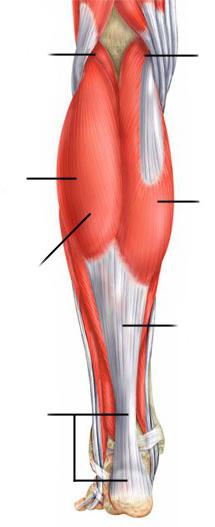
Among the muscles of the lower leg, the anterior, lateral and posterior muscle groups are distinguished. The anterior group mainly includes the extensors of the foot, the lateral group includes the flexors and pronators of the foot, and the posterior group includes the flexors and arch supports of the foot.
Rice. 135. Muscles of the lower leg (front view):
1 - long peroneal muscle; 2 - medial head of the gastrocnemius muscle; 3 - anterior tibial muscle; 4 - soleus muscle; 5 - short peroneal muscle; 6 - long extensor of the fingers; 7 - upper extensor retinaculum; 8 - tendon of the anterior tibial muscle; 9 - lower extensor retinaculum
front group
The anterior tibialis muscle (m. tibialis anterior) (Fig. 90, 135, 142, 146) unbends and adducts the foot, raising its medial edge. A long, narrow, superficial muscle originating on the lateral condyle of the tibia and the interosseous membrane. The attachment site is located on the plantar surface of the medial sphenoid bone and on the base of the I metatarsal bone. There is also a dry bag of the anterior tibial muscle (bursa subtendinea m. tibialis anterioris).
The long extensor digitorum longus (Fig. 90, 135, 141, 142, 146) unbends II-V fingers, as well as the foot, raising its lateral (outer) edge along with the third peroneal muscle. The muscle starts from the upper epiphysis of the tibia, the head and anterior edge of the fibula and the interosseous membrane. The muscle passes into a long narrow tendon, which divides into five thin individual tendons. Four of them are attached to the back of the II-IV fingers in such a way that the middle bundles of tendons are attached to the base of the middle phalanx, and the lateral ones - to the base of the distal phalanx. The fifth tendon attaches to the base of the fifth metatarsal.
The long extensor of the thumb (m. extensor hallucis longus) (Fig. 136) unbends the thumb, as well as the foot itself, raising its medial edge. Partially covered by the two previous muscles, located between them. The point of its beginning is the lower part of the medial surface of the body of the fibula, and the attachment point is the base of the distal phalanx. Part of the tendon bundles fuses with the base of the proximal phalanx.
Lateral group
The long peroneal muscle (m. peroneus longus) (Fig. 135, 137, 138, 139, 144, 146) abducts and flexes the foot, lowering its medial edge. Located on the lateral surface of the leg. The muscle starts from the head and upper body of the fibula and is attached to the medial sphenoid bone and the base of the I-II metatarsal bones.
Short peroneal muscle (m. peroneus brevis) (Fig. 135, 136, 138, 139, 140) removes and flexes the foot, raising its lateral edge. This long and thin muscle is located on the outer surface of the fibula. It is covered by the long peroneal muscle. The point of its beginning is located on the lower half of the lateral surface of the body of the fibula and the intermuscular septum. The place of attachment is the tuberosity of the V metatarsal bone.
back group
The back group includes two muscle groups.
Surface layer
The triceps muscle of the lower leg (m. triceps surae) flexes the lower leg at the knee joint, flexes and rotates the foot outward. With a fixed position of the foot, it pulls the lower leg and thigh backwards. The muscle consists of the superficial gastrocnemius muscle and the deep soleus muscle. Calf muscle (m. gastrocnemius) (Fig. 90, 132, 133, 134, 135, 137, 138, 146) has two heads. The medial head (caput mediale) starts from the medial epicondyle of the femur, and the lateral head (caput laterale) - from the lateral epicondyle. Both heads are connected into a common tendon and attached to the calcaneal tuber. The soleus muscle (m. soleus) (Fig. 90, 135, 137, 138, 139, 146) is covered by the gastrocnemius muscle, starts from the head and upper third of the posterior surface of the body of the fibula and from the line of the soleus muscle of the tibia. The muscle is attached on the calcaneal tubercle, growing together with the tendon of the gastrocnemius muscle. The common tendon in the lower third of the lower leg forms the calcaneal tendon (tendo calcaneus) (Fig. 137, 138), the so-called Achilles tendon. The mucous bag of the calcaneal tendon (bursa tendinis calcanei) is also located here.
The plantar muscle (m. plantaris) (Fig. 134, 137, 138) stretches the capsule of the knee joint during flexion and rotation of the lower leg. The muscle is rudimentary and unstable, has a spindle shape. Its point of origin is located on the lateral condyle of the femur and the bag of the knee joint, and the place of attachment is on the calcaneus.

Rice. 136. Muscles of the lower leg and foot (front view):
1 - articular muscle of the knee; 2 - square muscle of the thigh; 3 - short peroneal muscle; 4 - long extensor of the big toe; 5 - short extensor big toe; 6 - tendon of the long extensor of the big toe; 7 - short extensor of the fingers

Rice. 137. Muscles of the lower leg (back view):
1 - plantar muscle; 2 - gastrocnemius muscle: a) medial head, b) lateral head; 3 - soleus muscle; 4 - fascia of the lower leg; 5 - tendon of the posterior tibial muscle; 7 - tendon of the long flexor of the fingers; 8 - calcaneal tendon (Achilles tendon)

Rice. 138. Muscles of the lower leg (back view):
1 - plantar muscle; 2 - popliteal muscle; 3 - soleus muscle; 4 - tendon of the plantar muscle; 5 - gastrocnemius muscle: a) medial head, b) lateral head; 6 - tendon of the long peroneal muscle; 7 - tendon of the posterior tibial muscle; 8 - short peroneal muscle; 9 - tendon of the long flexor of the fingers; 10 - calcaneal tendon (Achilles tendon)

Rice. 139. Muscles of the lower leg (back view):
1 - popliteal muscle; 2 - soleus muscle; 4 - long peroneal muscle; 5 - long finger flexor; 6 - long flexor of the thumb; 7 - short peroneal muscle; 8 - flexor retainer; 9 - upper retainer of the long and short peroneal muscles

Rice. 140. Muscles of the lower leg and foot (rear view):
1 - popliteal muscle; 2 - short peroneal muscle; 3 - posterior tibial muscle; 4 - short flexor of the big toe; 5 - short flexor of the little toe of the foot; 6 - tendons of the long flexor of the fingers; 7 - interosseous muscles
deep layer
The popliteal muscle (m. popliteus) (Fig. 138, 139, 140) flexes the lower leg, rotating it inward and pulling the capsule of the knee joint. A short flat muscle, located on the posterior surface of the capsule of the knee joint, starts from it and from the lateral condyle of the femur, and is attached to the posterior surface of the body of the tibia.
The long flexor of the fingers (m. flexor digitorum longus) (Fig. 90, 137, 138, 139, 140, 143, 146) flexes the distal phalanges of the II-V fingers and takes part in the rotation of the foot outward, raising its medial edge. It is located on the posterior surface of the tibia, starts from the middle third of the posterior surface of the body of the tibia and from the deep sheet of the fascia of the leg. The tendon of the muscle is divided into four tendons, which are attached to the base of the distal phalanges of the II-V fingers.
The long flexor of the thumb (m. flexor hallucis longus) (Fig. 139, 143, 146) flexes the thumb, takes part in the flexion of the II-V fingers due to fibrous bundles, which are a continuation of the tendon, and also flexes and rotates the foot. The muscle originates from the lower two-thirds of the posterior surface of the body of the fibula and from the interosseous membrane, and is attached at the base of the distal phalanx of the thumb.
The tibialis posterior muscle (m. tibialis posterior) (Fig. 137, 138, 139, 140, 146) flexes and adducts the foot, rotating it outward. It is located on the interosseous membrane between the two previous muscles and is partially covered by the long flexor of the thumb. Its point of origin is on the posterior surfaces of the bodies of the tibia and fibula, and the place of attachment is on the sphenoid bones of the foot and the tuberosity of the scaphoid.
5th metatarsal. long muscle outperforms the short one by more than 2 times. Both muscles act as evertors and pronators. When walking during the transfer period, there is an increase in muscle activity before contact with the support. In the phase of the front push, the peroneal muscle pronates the foot. Muscle contraction leads to eversion of the hindfoot and prevents the heel from turning inward. At the end of the support period, before and during the back push, there is an increase in muscle activity. Their contraction causes stabilization of the midfoot and ray 1 joints. Tension in the tendon of the long peroneal muscle, which runs next to the cuboid bone, helps stabilize the calcaneocuboid joint, which makes it easier to push the foot off the support. Both peroneal muscles control the stability of the body in walking and standing. When standing on one leg, the work of the peroneal muscle allows you to maintain balance in the frontal plane. When the stability of the body is violated, an increase in muscle activity occurs. The role of the long peroneal muscle in walking depends on the speed of movement. When walking slows down, the role of the muscle in maintaining balance increases, and when walking accelerates, its role decreases.
Posterior leg muscles
Triceps muscle of the leg. The muscle has three heads. The inner and outer superficial heads start from the medial and lateral condyles of the thigh and form the gastrocnemius muscle. The deep head, or soleus muscle, starts from the top of the tibia and fibula. The triceps muscle is the main flexor of the foot at the ankle joint. When walking in the phase of the front push, the muscle contracts in an eccentric mode. It limits ankle extension, stabilizes it, and interacts with the quadriceps femoris muscle to extend the knee joint. In the rear push phase, the muscle exhibits maximum concentric activity. Its contraction provides flexion of the foot, repulsion by the foot from the support and lifting the body up.
Tibialis posterior. It comes from the interosseous membrane of the lower leg, passes under the medial condyle, attaches to the tuberosity of the navicular bone and the plantar surface of all the sphenoid bones. The muscle performs flexion and inversion of the foot in the subtalar joint and adduction of the midfoot, which provides supination of the foot. The muscle performs the function of the main dynamic stabilizer of the arch of the foot. It supports the arch of the foot and prevents it from lowering. The work of the muscle determines the position and ratio of the bones in the joints of the hind and middle sections of the foot. When walking in the forward thrust phase, there is eccentric muscle activity that counteracts the pronation of the foot. During the rear thrust phase, the muscle contracts concentrically, resulting in inversion of the foot, giving the joints of the foot the rigidity needed to push off the ground.
Muscle balance
The strength of the muscle is determined by its moment relative to the axis around which the segment rotates due to the action of this force. The moment of force is equal to the product of the magnitude of the force by the magnitude of its arm. A shoulder is a perpendicular dropped from the axis to the line of action of the force. The muscles of the lower leg, which carry out movement and stabilization in the joints of the foot, are located around the tibia. The tibialis anterior muscle is in front, the peroneal muscles are on the outside, the triceps muscle is on the back, and the tibialis posterior muscle is on the inside. Individual muscles have different strengths. The strongest are the muscles of the back group of the lower leg. They provide half the propulsion force when walking. The flexors of the foot are 3 times stronger than the extensors. The soleus muscle is one and a half times stronger than both heads of the gastrocnemius muscle. The ratio of muscle-inverter strength to evertors is 1:1. The forces that develop individual muscles located on opposite sides of the axis of the lower leg and having different masses are balanced with each other. The moments of some muscles are balanced by the moments of other muscles. The activity of all muscles is coordinated. Balanced work muscles provides leg support, absorption of shock load and repulsion of 6 tons of support in walking, maintaining the stability of the whole body.
The lack of work of some muscles leads to the predominance of traction of other muscles, resulting in muscle imbalance in several planes. The movement of the foot in the sagittal plane is controlled by the anterior tibialis and triceps muscles. In the absence of their contraction, the foot is in a state of free hanging under the influence of its own weight. With the weakening of the extensors of the foot, the traction of the flexors predominates, as a result of which the foot assumes the position of the equinus. With the weakening of the flexors, the predominance of the extensor muscles occurs, resulting in the formation of a calcaneal installation. In the frontal plane, the movements of the foot are controlled by the posterior tibial and peroneal muscles. The posterior tibialis inverts at the subtalar joint and adducts at the midtarsal joint. The peroneal muscles produce eversion at the subtalar joint and abduction at the midtarsal joint. Simultaneous contraction of both muscles leads to stabilization of the mid-tarsal joint.
Load on the bones of the leg
In the standing position, the tibia and fibula experience unequal stress. 85% of the weight from the block of the talus is transferred to the plafond of the tibia, 15% of the weight is transferred to the fibula through the tibiofibular syndesmosis and the talo-peroneal articulation. During walking, external and internal forces act on the bones of the lower leg. The external force is due to the influence of factors such as the reaction of the ground, the surface of the ground and the design of the shoe. Internal strength depends on the structure of the limb, bone elasticity, bone density and muscle tension. During walking, the tibia is simultaneously subjected to the action of the following forces: compression, bending, torsion, displacement, the main of which is the compression force. Compression and bending forces are determined by the magnitude and acceleration of the support reaction, as well as muscle contraction. The displacement load depends on the angle of inclination of the lower leg relative to the support, which changes during the support period. The torsion force is related to the external and internal rotations of the lower leg on the foot. Contraction of the calf muscles increases the compression force and decreases the displacement force. The simultaneous action of all forces during walking causes, first, during the front push, an increase in the compression force along the posterior surface of the bone, and then, during the back push, an increase in the compression force along the anterior surface of the bone.
Individual parameters of the mechanical load affect the structure of the bone. The magnitude of the load determines the structure of the bone trabeculae. An increase in load leads to an increase in the mass and number of trabeculae. The duration of the load affects the thickness of the bone cortical. An increase in the duration of activity leads to an increase in the area of the cortical layer of the bone.
The bone resists the force acting on it, which depends on the point of application of the force. Under the action of a compressive force along the center of the tibia, it undergoes compression throughout the entire ^ section. The resistance of the bone depends on the strength of the material. When loaded in the center, the strength of the material is used to the maximum. With an eccentric application of a compression force relative to the axis of the bone, the bearing capacity of the bone is determined by the moment of the internal pair of forces, the shoulder of which depends on the distribution of the strength of the material over the bone. There is an indirect relationship. With an eccentric application of force, the strength of the material has a lesser effect on the resistance of the bone; it does not affect the compression of the bone along the axis, when it experiences a bend, and the axis of the bone becomes curvilinear. The amount of bending depends on the elasticity of the bone and on the point of application of the force. The bone structures on the convex side experience tension, and on the concave side - compression. Between the compressed and stretched areas there is a neutral layer, which falls on the area of the bone marrow canal of the bone diaphysis. The cross-sections of the bone, which are parallel under conditions of a flat bone, form an angle with each other when bent.