Like the muscles of the thigh and pelvic girdle, they are relatively strongly developed. Their auxiliary apparatuses are sufficiently developed, which is determined by their load due to upright posture, the musculoskeletal function of the lower limb. Having an extensive origin on the bones, intermuscular septa and fascia of the lower leg, the lower leg muscles act on the knee, ankle and foot joints.
Distinguish between anterior, posterior and lateral group leg muscles. The muscles of the anterior group include the anterior tibial muscle, the long extensor of the fingers, the long extensor thumb; to the back - the triceps muscle of the lower leg (consisting of the gastrocnemius and soleus muscles), the plantar muscle, the popliteal muscle, the long flexor of the fingers, the long flexor of the large toe, tibialis posterior; to the lateral - long and short peroneal muscles.
Anterior leg muscles
Tibialis anterior located on the anterior surface of the lower leg, starts from the lateral condyle and the upper half of the lateral surface of the body of the tibia, the adjacent part of the interosseous membrane and from the fascia of the lower leg. At the level of the distal third of the lower leg, the muscle bundles pass into a long tendon, which successively passes under the upper and lower extensor retinaculums (tendons) anterior to the ankle joint, flexes the medial edge of the foot and attaches to the plantar surface of the medial sphenoid bone and to the base of the 1st metatarsal bone. The muscle unbends the foot in the ankle joint, simultaneously raises the medial edge of the foot and turns outward (supination), strengthens the longitudinal arch of the foot; with a fixed foot, tilts the lower leg forward; helps to keep the leg in a vertical position.
Long finger extensor- feathery muscle. It starts from the lateral condyle of the tibia, the anterior surface of the body of the fibula, from the upper third of the interosseous membrane, fascia and the anterior intermuscular septum of the leg.
Heading to the rear of the foot, the muscle sequentially passes behind the upper and lower extensor retinaculums (tendons). At the level of the ankle joint, it is divided into four tendons, which are enclosed in a synovial sheath common to them. Each tendon is attached to the base of the middle and distal phalanges of fingers 2-5.
A small bundle is separated from the lower part of the muscle - the third peroneal muscle, the tendon of which is attached to the base of the 5th metatarsal bone. The muscle extends 2-5 fingers in the metatarsophalangeal joints, as well as the foot in the ankle joint. The third peroneal muscle raises the lateral edge of the foot. With a strengthened foot, similarly to the anterior tibial muscle, the long extensor of the fingers holds the lower leg in a vertical position.
Long extensor thumb foot is located between the anterior tibial muscle medially and long extensor fingers laterally, partially covered by them in front. It starts from the middle third of the anterior surface of the fibula, the interosseous membrane of the leg. The tendon of the muscle passes down to the dorsum of the foot under the superior and inferior extensor retinaculums (tendons) in a separate synovial sheath and attaches to the distal phalanx of the big toe. Separate tendon bundles can also attach to the proximal phalanx. The muscle extends the big toe; also involved in the extension of the foot in the ankle joint.
 Triceps muscle of the leg consists of two muscles - the gastrocnemius muscle, which is located superficially, and the soleus muscle, hidden under the gastrocnemius. The gastrocnemius muscle belongs to the biarticular muscles, it passes through two joints - the knee and ankle, while the soleus muscle is single-joint - it passes only through the ankle joint.
Calf muscle has two heads) - medial and lateral, the surface layers of which are represented by strong tendon bundles. The lateral head begins on the outer surface of the lower epiphysis of the femur above the lateral condyle; medial head - on the medial condyle of the thigh. Under each of the heads of the gastrocnemius muscle is a synovial bag. Between the lateral head and the capsule of the knee joint, there is a lateral podshezhinous bag of the gastrocnemius muscle. Between the medial head and the joint capsule lies the medial gastrocnemius bursa. Both bags, as a rule, communicate with the cavity of the knee joint.
In the middle of the lower leg, both heads of the gastrocnemius muscle pass into a thick tendon, which narrows downward and merges with the tendon of the soleus muscle, forming the calcaneal (Achilles) tendon, which is attached to the calcaneal tuberosity. Between the tendon and the bone there is a synovial bag - a bag of the calcaneal tendon.
soleus muscle thick, flat, lying in front of the gastrocnemius muscle. In front of it are the muscles of the deep layer. The soleus muscle has an extensive origin on the posterior surface of the tibia and from the tendon arch that extends between the tibia and fibula. The muscle of the pennate structure passes into a flat tendon, which is involved in the formation of the calcaneal tendon. The triceps muscle of the lower leg flexes the lower leg and foot (plantar flexion); with a fixed foot, it holds the lower leg on the talus, preventing it from tipping forward.
plantar muscle inconsistent, with a small abdomen and a long thin tendon. It originates on the lateral epicondyle of the thigh and from the oblique popliteal ligament. The tendon of this muscle passes between the gastrocnemius and soleus muscles, is adjacent to the medial edge of the calcaneal tendon, with which it is attached to the calcaneal tuberosity. The muscle stretches the capsule of the knee joint, is involved in the flexion of the lower leg and foot.
Hamstring lies deep in the popliteal fossa. It begins with a thick tendon from the outer surface of the lateral condyle of the thigh (below the attachment of the peroneal collateral ligament). The muscle is adjacent to the posterior surface of the knee joint and passes under the arcuate popliteal ligament, from which its medial bundles begin. It is attached to a triangular platform on the posterior surface of the tibia, above the line of the soleus muscle. The muscle flexes the lower leg, turning it inwards; stretches the capsule of the knee joint, protecting the synovial membrane from infringement.
Long finger flexor- bifid muscle. It begins in fleshy bundles on the posterior surface of the body of the tibia below the line of the soleus muscle, from the fascia of the lower leg and from the posterior intermuscular septum of the lower leg. They are located behind and medial to the posterior tibial muscle.
The tendon of the long flexor of the fingers goes down, crosses the tendon of the posterior tibial muscle from behind and from the lateral side. Further, the tendon of the muscle passes to the sole of the foot behind the medial malleolus under the retinaculum (tendons) of the flexors in a separate synovial sheath (between the tendons of the posterior tibial muscle medially and the long flexor of the thumb laterally). Then the tendon goes around the back and bottom of the support of the talus, located above the short flexor of the fingers, is divided into four separate tendons, which are attached to the distal phalanges of 2-5 fingers, pre-perforating the tendons of the short flexor of the fingers (similar to the tendons of the deep flexor of the fingers on the hand). The muscle flexes the distal phalanges of 2-5 fingers; flexes the foot, turning it outward.
flexor thumb longus feet - bipennate muscle. It starts from the lower two-thirds of the body of the fibula, the interosseous membrane, the posterior intermuscular septum of the leg. It is located laterally and behind the tibialis posterior muscle. The flexor hallucis longus tendon passes under the flexor retinaculum (tendons) behind the medial malleolus and lateral to the flexor hallucis longus tendon in a separate synovial sheath. Further, the tendon of the long flexor of the big toe lies in the groove of the same name on the posterior process of the talus, passing forward under the support of the talus. Upon reaching the plantar surface of the big toe, the tendon attaches to the distal phalanx of the big toe. On its way on the foot, this tendon crosses with the tendon of the long flexor of the fingers (lies under it). Throughout the plantar surface of the I metatarsal bone, the tendon of the long flexor of the big toe lies between the medial and lateral abdomens of the short flexor of the big toe. The muscle flexes the big toe, is involved in flexion, supination and adduction of the foot; strengthens the longitudinal arch of the foot.
Tibialis posterior located deep on the back of the leg, between the long flexor of the fingers (medially) and the long flexor of the big toe (laterally). It starts on the posterior surface of the body of the fibula (between the medial crest and interosseous margin), from the lower surface of the lateral condyle and the upper two-thirds of the body of the tibia (below the line of the soleus muscle) and the interosseous membrane of the leg. The muscle continues into a strong tendon that lies in a groove on the posterior surface of the medial malleolus in front of the tendon of the long flexor of the fingers by the flexor tendon retinaculum. Moving to the plantar surface, the tendon is attached to the tuberosity of the navicular bone, to all three sphenoid bones, and also to the base of the 4th (sometimes 5th) metatarsal bone. The muscle flexes the foot (plantar flexion), adducts it, and supinates.
Lateral leg muscle group
The lateral group is represented by the long and short peroneal muscles, which are located on the lateral surface of the lower leg under the fascia plate, between the anterior and posterior intermuscular septa.
Peroneus longus muscle, two-pinnate, lies superficially. It starts from the head and the upper two-thirds of the lateral surface of the fibula, from the lateral condyle of the tibia, the fascia of the lower leg and from the intermuscular septa of the lower leg. At the level of the ankle joint, the tendon of the muscle, bending around the lateral ankle from behind, passes first under the upper retinaculum (tendons) of the peroneal muscles in the common synovial sheath with the tendon of the short peroneal muscle, and then in the groove on the calcaneus under the lower retinaculum (tendons) of the peroneal muscles. On the sole, the tendon of the long peroneal muscle runs obliquely forward and medially, lies in the groove of the same name in the cuboid bone in a separate (own) synovial sheath; attached to the base of the 1st and 2nd metatarsal bones and to the medial sphenoid bone.
At points where the tendon changes direction (behind the lateral malleolus and on the cuboid bone), it usually thickens due to the fibrocartilage or sesamoid bone that forms in its thickness. The muscle flexes the foot, raises its lateral edge (pronation), strengthens the transverse and longitudinal arches of the foot.
Peroneus brevis bipinnate, starts from the lower two-thirds of the lateral surface of the fibula and from the intermuscular septa of the lower leg. The tendon of the muscle passes to the foot behind the lateral malleolus, lying in the common synovial sheath together with the tendon of the long peroneal muscle under the retinaculum (tendons) of the peroneal muscles. At the lower edge of this retainer, the tendon of the short peroneal muscle turns forward and passes along the outer side of the calcaneus under the fibular block to the place of attachment at the base of the 5th metatarsal. The muscle raises the lateral edge of the foot; prevents the foot from turning with the sole inside; flexes the foot (plantar flexion).
The posterior muscle group of the leg.
Surface layer (calf muscles):
M. triceps surae, triceps leg muscle, forms the main mass of the calf elevation. It consists of two muscles - m. gastrocnemius, located superficially, and m. soleus, lying under it; both muscles below have one common tendon.
- M. gastrocnemius, gastrocnemius, originates from facies poplitea femur behind both condyles with two heads, which, with their tendon origin, fuse with the capsule of the knee joint. The heads pass into the tendon, which, merging with the tendon m. soleus, continues into the massive Achilles tendon, tendo calcaneus (Achillis), which is attached to the posterior surface of the calcaneal tubercle. At the point of attachment between the tendon and the bone, there is a very permanent synovial bag, bursa tendinis calcanei (Achillis).
- M. soleus, soleus muscle, thick and fleshy. Lies under calf muscle, occupying a large extent on the bones of the lower leg. The line of its beginning is located on the head and on the upper third of the posterior surface of the fibula and descends along the tibia almost to the border of the middle third of the lower leg from the bottom. In the place where the muscle is thrown from the fibula to the tibia, a tendon arch is formed, arcus tendineus m. solei, under which the popliteal artery and n. tibialis. Tendon sprain m. soleus merges with the Achilles tendon.

M. plantaris, plantar muscle. It originates from the facies poplitea above the lateral condyle of the thigh and from the capsule of the knee joint, soon passes into a very long and thin tendon that stretches in front of m. gastrocnemius and is attached at the calcaneal tubercle. This muscle undergoes reduction and in humans is a rudimentary formation, as a result of which it may be absent. Function. All muscles m. triceps surae (including m. plantaris) produces flexion in the ankle joint both with a free leg and with support on the end of the foot. Since the line of traction of the muscle passes medially to the axis of the subtalar joint, it also makes the adduction of the foot and supination. When standing, the triceps surae (especially m. soleus) prevents the body from tipping forward at the ankle joint. The muscle has to work mainly when burdened with the weight of the whole body, and therefore it is strong and has a large physiological diameter; m. gastrocnemius, as a biarticular muscle, can also flex the knee with a strengthened lower leg and foot. (Inn. m. triceps surae and m. plantaris - L5-S2. N. tibialis.) The deep layer, separated from the superficial deep fascia of the lower leg, is composed of three flexors that oppose three similar extensors lying on the anterior surface of the lower leg.

M. flexor digitorum longus, long finger flexor, the most medial of the muscles of the deep layer. It lies on the posterior surface of the tibia, from which it originates. The tendon of the muscle descends behind the medial malleolus, in the middle of the sole it is divided into four secondary tendons, which go to the four fingers II-V, pierce like a deep flexor on the tendon brush m. flexor digitorum brevis and are attached to the distal phalanges. The function in terms of finger flexion is small; the muscle mainly acts on the foot as a whole, producing flexion and supination of it with a free leg. She also, along with m. triceps surae is involved in putting the foot on the toe (walking on tiptoe). When standing, the muscle actively contributes to the strengthening of the arch of the foot in the longitudinal direction. When walking, he presses his toes to the ground. (Inn. L5-S1. N. tibialis.)

M. tibialis posterior, posterior tibial muscle, occupies the space between the bones of the lower leg, lying on the interosseous membrane and partly on the tibia and fibula. From these places, the muscle receives its initial fibers, then bends around the medial malleolus with its tendon and, having reached the sole, is attached to the tuberositas ossis navicularis, and then in several bundles to the three sphenoid bones and the bases of the II-IV metatarsal bones. Function. Bends the foot and brings it together with m. tibialis anterior. Together with other muscles that are also attached to the medial edge of the foot (m. tibialis anterior et m. peroneus longus), m. tibialis posterior forms, as it were, a stirrup that strengthens the arch of the foot; stretching his tendon through the lig. calcaneonavicular, the muscle supports the head of the talus together with this ligament. (Inn. L5-S1. N. tibialis.)
M. flexor hallucis longus, long flexor of the big toe, the most lateral of the muscles of the deep layer. Lies on the posterior surface of the fibula, from which it originates; the tendon runs in a groove on the processus posterior of the talus, fits under the sustentaculum tali to the thumb, where it attaches to its distal phalanx. Function. Flexes the thumb, and also due to the possible connection with the tendon m. flexor digitorum longus can act in the same sense on Pi even III and IV fingers. Like the rest of the posterior muscles of the lower leg, m. flexor hallucis longus produces flexion, adduction and supination of the foot and strengthens the arch of the foot in the anteroposterior! direction. (Inn. L5-S2. N. tibialis.)
Anterior leg muscles
Anterior tibial muscle (m. tibialis anterior) (Fig. 197) is located on the anterior surface of the lower leg. It has a wide origin from the lateral upper third of the tibia, the fascia of the lower leg and the interosseous membrane. It passes near the anterior tibial crest under the retinaculum mm. extensorum superius et inferius in the fibrous canal and exits on the medial edge of the foot, where the tendon is attached to the plantar surface of the I sphenoid and metatarsal bones.
Function. Extends at the ankle joint and supinates the foot.
The long extensor of the first finger (m. extensor hallucis longus) (Fig. 197) is located lateral to m. tibialis anterior. It starts from the fibula and the interosseous membrane. It comes out between the anterior tibial muscle and the long extensor of the fingers. The tendon passes through the fibrous channel under the retinaculum mm. extensorum superius et inferius, ends at the base of the distal phalanx of the first finger.
Innervation: n. peroneus profundus (LIV-SI).
Function. Corresponds to the name of the muscle. In addition, the muscle is involved in the extension of the foot in the ankle joint.
197. Muscles of the lower leg and foot. 1 - tendo m. sartorius; 2 - tibia; 3 - m. gastrocnemius; 4 - m. soleus; 5 - m. tibialis anterior; 6 - tendo m. extensoris hallucis longi; 7 - tendo m. extensoris digitrum longi; 8 - retinaculum mm. extens6rum inferius; 9 - m. peroneus brevis; 10 - m. peroneus longus; 11-lig. patellae; 12 - tractus iliotibialis.
The long extensor of the fingers (m. extensor digitorum longus) is located lateral to m. tibialis anterior, covers the long extensor of the first finger. It starts from the upper third of the tibia, fibula, membrana interossea and fascia of the leg. The muscle is separated from the anterior tibialis muscle intermuscular septum. Forms a tendon that runs in the fibrous sheath under the retinaculum mm. extensorum inferius. Upon reaching the foot, the tendon is divided into 4 tendons, which are attached to the aponeurotic plate of the rear of the II-V fingers.
Innervation: n. peroneus profundus (LIV-SI).
Function. Unbends fingers II-IV, penetrates the outer edge of the foot together with the third peroneal muscle.
The third peroneal muscle (m. peroneus tertius) represents the fifth part of the long extensor of the fingers. This muscle is unstable (8.2%). It is attached to the fascia of the lateral part of the rear of the foot and to the fifth metatarsal bone.
The muscle is a derivative of the permanent muscle m existing in monkeys. peroneus parvus.
Innervation: n. peroneus profundus (LIV-SI).
Function. Unbends the foot at the ankle joint, raises the lateral edge of the foot.

198. Muscles of the lower leg and foot from the lateral side.
1 - m. extensor digitorum longus;
2 - m. extensor digitorum brevis;
3 - malleolus lateralis;
4 - m. peroneus brevis;
5 - m. peroneus longus;
6 - m. soleus;
7 - m. gastrocnemius;
8 - m. biceps femoris;
9 - tractus iliotibialis.
Lateral muscles of the leg
Long peroneal muscle (m. peroneus longus) (Fig. 198) occupies the lateral region of the lower leg, separated by an intermuscular septum from the long extensor of the fingers and m. soleus. It starts in two bundles from the head and body of the upper part of the fibula, the lateral tibial condyle and the fascia of the leg. The superficial peroneal nerve passes between the heads in the canalis musculoperoneus. The tendon arises above the lateral malleolus and passes under the retinaculum mm. peroneorum superius in the fibrous canal along with the tendon of the short peroneal muscle, bending around the lateral malleolus. Having reached the rear of the foot, the tendon penetrates the sole along the sulcus ossis cuboidei, where it reaches the medial edge of the foot, attaching to the I metatarsal and I cuneiform bones. On the sole, the tendon passes in the bone-fibrous canal.
Function. Flexes the foot at the ankle joint, raises the lateral edge of the foot.
The short peroneal muscle (m. peroneus brevis) lies under the previous one, shorter than it by a third. It starts from the fibula and intermuscular septa. The tendon of the muscle lies first in front of the long peroneal muscle, and then behind it, passes in the common fibrous canal, attaches to the tuberosity of the fifth metatarsal bone.
Innervation: n. peroneus superficial (LV-SI).
Function: Flexes and pronates the foot.
Back muscles of the leg
The triceps muscle of the leg (m. triceps surae) has three heads. The gastrocnemius muscle (m. gastrocnemius) starts from the areas above the lateral and medial condyles of the thigh with two heads, forming the lower border of the fossa poplitea, and also, together with the posterior wall of the articular capsule, limits the entrance to the canalis cruropopliteus; the soleus muscle (m. soleus) is covered by the gastrocnemius muscle. Starting from the linea poplitea tibiae, the head of the fibula and the tendon arch stretched between the bones of the lower leg, it connects below into a single powerful calcaneal tendon of the triceps muscle of the lower leg - tendo calcaneus (Achillis), attached to the calcaneal tubercle. Between the tendon and the calcaneal tuber there is a mucous bag.
Innervation: n. tibialis (LIV-SII).
Function. Flexes the foot at the ankle joint. When walking and running pushes the foot off the ground.
The plantar muscle (m. plantaris) starts from the area above the condyle of the thigh and the capsule of the knee joint. Then a thin tendon penetrates between the gastrocnemius and soleus muscles and is woven into the tendon of the triceps muscle of the lower leg.
Innervation and function. Same as the calf muscle.
The long flexor of the fingers (m. flexor digitorum longus) is located on the medial surface of the lower leg. It starts from the middle third of the posterior surface of the tibia and the deep fascia of the lower leg. The tendon reaches the medial malleolus and under the retinaculum mm. flexorum in the fibrous canal passes to the foot between the tendons m. tibialis posterior and m. flexor hallucis longus. On the foot it crosses with the tendon m. flexor hallucis longus, receiving from it a fibrous bundle of fibers. From the long flexor of the fingers also begins part of the muscle bundles m. quadratus plantae. Then the long flexor of the fingers is divided into four tendons, which, piercing the tendon of the short flexor of the fingers in the region of the phalanges, are attached to the base of the distal phalanges from II to V finger.
Innervation: n. tibialis (LV-SI).
Function. Bends the fingers, on which the foot rests when walking, and the foot at the ankle joint.
The tibialis posterior muscle (m. tibialis posterior) (Fig. 199) starts from the interosseous membrane and the bones of the lower leg of the entire posterior surface. The lower part is covered by the flexors of the fingers. The squamous tendon runs behind the medial malleolus and inserts on the tuberosity of the navicular and all of the cuneiform bones.
Function. Flexes at the ankle joint and supinates the foot, participates in maintaining its arches.
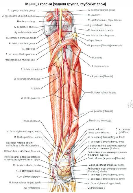
199. Muscles of the lower leg, rear view.
1 - m. gastrocnemius; 2 - m. soleus; 3 - m. tibialis posterior; 4 - m. flexor hallucis longus; 5 - m. peroneus longus; 6 - m. peroneus brevis; 7 - m. flexor digitorum longus; 8 - m. popliteus
The long flexor of the first finger (m. flexor hallucis longus) is a more massive muscle than the long flexor of the fingers and the posterior tibial muscle. It is located lateral to the previous muscles, bordering on the long and short peroneal muscles. It starts from the fibula and the intermuscular septum. Passes behind the medial malleolus and sustentaculum tali, surrounded by the synovial sheath in the fibrous canal. Attached to the distal phalanx of the first finger. Sesamoid bones are often found in the tendon.
Innervation: n. tibialis (LV-SII).
Function. Bends I finger, supports the inner arch of the foot. Due to the fibrous bundle that has entered the long flexor of the fingers, to some extent it helps to bend the other fingers.
The lower leg refers to the lower limb. It is located between the foot and the knee area. The lower leg is formed by means of two bones - small and tibial. They are surrounded by muscle fibers on three sides. The muscles of the lower leg, the anatomy of which will be discussed later, set the fingers and foot in motion.
Tibia
This element has an extension on the top edge. Condyles are formed in this area: lateral and medial. On top of them are the surfaces of the joints. They articulate with the condyles of the thigh. On the lateral segment, there is an articular surface on the outside, through which it is connected to the head in the fibula. The body of the tibial element looks like a trihedral prism. Its base is directed backwards and has 3 surfaces, respectively: back, outer and inner. There is an edge between the last two. It's called the front. In its upper part, it passes into the tuberosity of the tibia. This area is intended for fixation. In the lower part, the tibia has an extension, and on inner surface protrusion is present. It is oriented downward. This protrusion is called the medial malleolus. On the back side of the bone lies a rough segment of the soleus muscle. The articular surface is located on the distal epiphysis. It serves to connect with

Second element
The fibula is thin, long, located laterally. Its upper end has a thickening - the head. It connects to the tibia. The lower section of the element is also thickened and forms the lateral malleolus. She, like the head of the fibula, is oriented outwards and is well palpable.
Leg muscles: their location, functions
The fibers are located on three sides. Allocate different muscles shins. The front group performs extension of the foot and fingers, supination and adduction of the foot. This segment includes three types of fibers. The tibialis anterior muscle of the lower leg was formed first. The remaining fibers form the long extensors of the fingers and a separate one for the big toe on the foot. The posterior muscle group of the lower leg forms a greater number of fibers. In particular, there are long finger flexors and separately for the large, popliteal, triceps muscle of the lower leg. There are also tibial fibers here. The outer group includes the short and long peroneal muscles of the lower leg. These fibers flex, penetrate, and abduct the foot.
Tibial segment
This anterior muscle of the lower leg starts from the bone of the same name, its outer surface, fascia and interosseous membrane. They are directed downward. The fibers pass under two ligaments. They are located in the area and ankles. These areas - the upper and lower retainers of the extensor tendons - are represented by places of thickening of the fascia of the foot and lower leg. The site of attachment of the fibers is the sphenoid medial and the base of the metatarsal (first) bone. The muscle is quite well palpable along its entire length, especially in the area of transition to the foot. In this place, her tendon protrudes during extension. The task of this leg muscle is the supination of the foot.

Finger extensor (long)
It runs from the anterior muscle outwards in the upper region of the lower leg. Its fibers begin from the head and marginal sections of the tibia, fascia and interosseous membrane. The extensor, passing to the foot, is divided into five tendons. Four are attached to the distal (from the second to the fifth), the last - to the base of the 5th metatarsal. The task of the extensor, acting as a multi-joint muscle of the lower leg, is not only to coordinate the extension of the fingers, but also the foot. Due to the fact that one tendon is fixed at its edge, the fibers also penetrate the area somewhat.
Extensors of the thumbs
The fibers begin in the region of the lower leg from the interosseous membrane and the inner part of the fibula. The extensors have less strength than the segments described above. The site of attachment of this is the distal phalanges in thumbs. These muscles of the lower leg not only carry out their extension, but also the feet, also contributing to their supination.

Finger flexor (long)
It starts from the back of the tibia, passing under the medial malleolus to the foot. The channel for it is located under the retainer. Next, the muscle is divided into four segments. On the foot (plantar surface), fibers cross the tendon from the flexor (long) thumb. Then the square muscle of the sole joins them. Four formed tendons are fixed to the distal phalanges (at their base) of 2-5 fingers. The task of this muscle is, among other things, to flex and supinate the foot. The fibers of the square segment are attached to the tendon. Due to this, the action of the muscle is averaged. Lying under the medial malleolus and fanning out towards the phalanges, the long flexor also provokes some adduction of the fingers to the median surface of the body. By pulling square muscle tendons, this action is slightly reduced.
Triceps muscle of the leg
It runs along the back surface and has 3 heads. Two form the superficial area - the gastrocnemius muscle, from the third - deep - the fibers of the soleus segment depart. All heads are connected and form a common Achilles (calcaneal) tendon. It is attached to the tubercle of the corresponding bone. The gastrocnemius muscle starts from the femoral condyles: lateral and medial. The task of the two heads located in this area is twofold. They coordinate flexion at the knee joint and the foot at the ankle joint. The medial element descends slightly lower and is better developed than the lateral one. From the back side in the upper third of the tibia, the soleus muscle departs. It is also attached to the tendon arch located between the bones. The fibers pass somewhat lower and deeper than the gastrocnemius. They lie behind the subtalar and cause flexion of the foot. The triceps muscle can be felt under the skin. The calcaneal tendon protrudes posteriorly from the transverse axis in the ankle joint. Due to this, the triceps muscle has a large moment of rotation relative to this line. The heads of the gastrocnemius segment are involved in the formation of the rhomboid popliteal fossa. Its boundaries are: two-headed thigh muscle(outside and top), semimembranous fibers (inside and top), plantar and two heads of the gastrocnemius segment (bottom). The bottom in the fossa is formed by the capsule of the knee joint and the vessels and nerves that feed the foot and lower leg run through this area.

Flexor (long) thumb
This muscle of the posterior surface of the lower leg is characterized by the greatest strength. On the plantar side of the foot, fibers run between the heads from a short segment responsible for flexion of the big toe. The muscle starts from the back side (lower part) of the fibula and the intermuscular septum (back). The site of fixation is the plantar surface of the base of the distal phalanx in the thumb. Due to the fact that the tendon of the muscle partially passes into the element of the long flexor of the same name, it has some influence on the movements of 2-3 fingers. The presence on the surface of the sole of the metatarsophalangeal joint of 2 large sesamoid bone elements provides an increase in the moment of rotation of the fibers. The tasks of the segment include flexion of the entire foot and thumb.
Second division of tibial fibers
This posterior segment is located under the triceps muscle. The fibers start from the interosseous membrane and areas of the small and tibial bones adjacent to it. The site of attachment of the muscle is the tubercle of the navicular, the base of the metatarsal and all the wedge-shaped elements. The muscle lies under the medial malleolus and performs flexion of the foot, supination and adduction. A canal passes between the soleus and tibial fibers. It is presented in the form of a gap. It contains nerves and blood vessels.

Popliteal segment
It is formed by flat short fibers. The muscle adjoins directly to the knee joint from behind. The fibers originate from the femoral condyle (lateral), below the gastrocnemius segment, and the bursa of the knee joint. They pass down and are attached above the soleus muscle to the tibia. Because the fibers are partially attached to the joint capsule, they pull it posteriorly when flexed. The task of the muscle is pronation and flexion of the lower leg.
Long peroneal segment
This muscle has a feathery structure. It runs along the surface of the fibula. It starts from its head, the condyle of the tibial element, partly from the fascia. It is also attached to the 2-thirds region. outside fibula. When the muscle contracts, abduction, pronation, and flexion of the foot occur. The tendon of the long peroneal segment posteriorly and inferiorly bypasses the lateral malleolus. In the area of the heel bone there are ligaments - the upper and lower retainers. When moving to the plantar part of the foot, the tendon runs along the groove. It is located on the underside of the cuboid bone. The muscle reaches the inside of the foot.

Short peroneal fibers
The tendon of the segment wraps around the lateral malleolus behind and below. It is attached to the tubercle on the 5th metatarsal. The segment begins from the intermuscular septa and the outer part of the fibula. The task of the fibers is abduction, pronation and flexion of the foot.
Anterior tibial muscle (m.tibialis anterior) is located on the front side of the lower leg. It starts on the lateral condyle and the upper half of the lateral surface of the body of the tibia, as well as the adjacent part of the interosseous membrane and on the fascia of the leg. At the level of the distal third of the lower leg, the muscle bundles pass into a long tendon that runs under the upper and lower extensor tendon retinaculum, anterior to the ankle joint. Further, the tendon goes around the medial edge of the foot and is attached to the plantar surface of the medial sphenoid bone and the base of the first metatarsal bone.
Function: unbends the foot in the ankle joint, simultaneously raises the medial edge of the foot and turns it outward (supination), strengthens the longitudinal arch of the foot. With a fixed foot, tilts the lower leg forward; helps to keep the leg in a vertical position.
Blood supply: anterior tibial artery
The extensor digitorum longus (m.extensor digitorum longus) is a pinnate muscle that starts on the lateral condyle of the tibia, the anterior surface of the body of the fibula, on the upper third of the interosseous membrane, fascia, and anterior intermuscular septum of the leg. Heading to the rear of the foot, the muscle passes behind the upper and lower extensor tendon retainers. At the level of the ankle joint, the muscle is divided into 4 tendons, which are enclosed in a synovial sheath common to them. Each tendon is attached to the back of the base of the middle and distal phalanges of the II-V fingers.
A small bundle is separated from the lower part of the muscle, called third peroneal muscle(m.peroneus tertius), the tendon of which is attached to the base of the fifth metatarsal bone.
Function: unbends the II-V fingers in the metatarsophalangeal joints, as well as the foot in the ankle joint. The third peroneal muscle raises the lateral edge of the foot. With a strengthened foot, the long extensor of the fingers holds the lower leg in a vertical position.

Innervation: deep peroneal nerve (LIV-SI). Blood supply: anterior tibial artery.
The long extensor of the big toe (m.extensor hallucis longus) is located between the anterior tibial muscle medially and the long extensor of the fingers laterally; partially covered by them in front. It starts on the middle third of the anterior surface of the fibula, the interosseous membrane of the leg. The tendon of the muscle passes down to the dorsum of the foot under the superior and inferior extensor tendon retinaculum in a separate synovial sheath and attaches to the distal phalanx of the big toe. Separate tendon bundles can also attach to the proximal phalanx.
Function: unbends the big toe; also involved in the extension of the foot in the ankle joint.
Innervation: deep peroneal nerve (LIV-SI).
Blood supply: anterior tibial artery.
, , ,
Posterior leg muscles
The muscles of the posterior group form two layers - superficial and deep. The superficially lying triceps muscle of the lower leg is more strongly developed, which creates the roundness of the lower leg characteristic of a person. The deep layer is formed by a small popliteal muscle and 3 long muscles: long flexor of the fingers (located most medially), posterior tibial muscle (occupies intermediate position) and long flexor of the big toe (located laterally).

Superficial layer of the posterior leg muscles
The triceps muscle of the lower leg (m.triceps surae) consists of two muscles - the gastrocnemius muscle, which is located superficially, and the soleus muscle, hidden under the gastrocnemius. The gastrocnemius muscle is a biarticular muscle, it acts on two joints - the knee and ankle, while the soleus muscle is single-joint - it acts only on the ankle joint.
Calf muscle(m.gastrocnemius) has two heads: medial and lateral, the surface layers of which are represented by strong tendon bundles. The lateral head (caput laterale) begins on the outer surface of the lower epiphysis of the thigh above the lateral condyle. The medial head (caput mediate) begins on the medial condyle of the thigh. Under each head of the gastrocnemius muscle is a synovial bag. Between the lateral head and the capsule of the knee joint is located lateral tendon bursa of the gastrocnemius muscle(bursa subtendinea musculi gastrocnemii lateralis). Between the medial head and the joint capsule is medial gastrocnemius bursa(bursa subtendinea musculi gastrocnemii medialis). Both bags, as a rule, communicate with the cavity of the knee joint.
In the middle of the lower leg, both heads of the gastrocnemius muscle pass into a thick tendon, which narrows downward and merges with the tendon of the soleus muscle, forming the calcaneal (Achilles) tendon (tendo calcaneus, s.Achilli), which is attached to the calcaneal tuberosity. Between the tendon and the calcaneus there is a bag of the calcaneal (Achilles) tendon (bursa tendinis calcanei, s.Achillis).
soleus muscle(m.soleus) thick, flat, lies under the calf muscle. In front of it are the muscles of the deep layer. The soleus muscle has an extensive origin on the posterior surface of the tibia (on the line of the soleus muscle) and on the tendon arch (arcus tendineus musculi solei), which extends between the tibia and fibula. The soleus muscle has a pinnate structure, passes into a flat tendon, which is involved in the formation of the calcaneal tendon.
Function: triceps flexes the lower leg and foot (plantar flexion); with a fixed foot, it holds the lower leg on the talus, preventing it from tipping forward.
Innervation: tibial nerve (LIV-SI).
plantar muscle
(m.plantaris) fickle, has a small abdomen and a long thin tendon. It originates on the lateral epicondyle of the thigh and on the oblique popliteal ligament. The tendon of this muscle passes between the gastrocnemius and soleus muscles, is adjacent to the medial edge of the calcaneal tendon, with which it is attached to the calcaneal tuberosity.
Function: stretches the capsule of the knee joint, participates in flexion of the lower leg and foot.
Deep layer of the posterior leg muscles
The deep layer is formed by 4 muscles: popliteal, long flexor of the fingers, long flexor of the big toe and posterior tibial muscle, which are separated from the soleus muscle by a deep plate of the fascia of the lower leg.

The popliteal muscle (m.popliteus) lies deep in the popliteal fossa. It begins with a thick tendon on the outer surface of the lateral condyle of the thigh (below the attachment of the peroneal collateral ligament). The muscle is adjacent to the posterior surface of the joint capsule and is located below the arcuate popliteal ligament, on which its medial bundles begin. The muscle attaches to a triangular area on the posterior surface of the tibia, above the line of the soleus muscle.
Function: flexes the lower leg, turning it inwards; stretches the capsule of the knee joint, protecting the synovial membrane from infringement.
Innervation: tibial nerve (LIV-SII).
Blood supply: popliteal artery.
The long flexor of the fingers (m.flexor digitorum longus) has a two-pinnate structure, begins with fleshy bundles on the posterior surface of the body of the tibia below the line of the soleus muscle, as well as on the fascia and posterior intermuscular septum of the leg. It is located behind and medial to the posterior tibial muscle. The tendon of the long flexor of the fingers goes down, crosses behind and from the lateral side the tendon of the posterior tibial muscle. Further, the tendon of the muscle passes to the sole of the foot behind the medial malleolus under the retinaculum of the flexor tendons in a separate synovial sheath (between the tendons of the posterior tibial muscle medially and the long flexor of the thumb laterally). Then the tendon goes around behind and below the support of the talus. Located above the short flexor of the fingers, it is divided into 4 separate tendons, which are attached to the distal phalanges of the II-V fingers, having previously pierced the tendons of the short flexor of the fingers (like the tendons of the deep flexor of the fingers on the hand).
Function: bends the distal phalanges of the II-V fingers; flexes the foot, turning it outward.
Innervation: tibial nerve (LIV-SII).
Blood supply: posterior tibial artery.
Long flexor of the big toe
(m.flexor hallucus longus) - bipennate muscle, begins on the lower two-thirds of the body of the fibula, interosseous membrane, posterior intermuscular septum of the leg. It is located laterally and behind the tibialis posterior muscle. The flexor hallucis longus tendon passes under the flexor tendon retinaculum behind the medial malleolus and lateral to the flexor hallucis longus tendon in a separate synovial sheath. Further, the tendon of the long flexor of the big toe lies in the groove of the same name on the posterior process of the talus, passing forward under the support of the talus. Having reached the plantar surface of the big toe, the tendon of the long flexor of the big toe is attached to its distal phalanx. On its way on the foot, this tendon crosses with the tendon of the long flexor of the fingers (lies under it). Throughout the plantar surface of the I metatarsal bone, the tendon of the long flexor of the big toe lies between the medial and lateral abdomens of the short flexor of the big toe.
Function: flexes the big toe, participates in flexion (supination) and adduction of the foot; strengthens the longitudinal arch of the foot.
Innervation: tibial nerve (LIV-SII).
Blood supply: posterior tibial and peroneal arteries.
The posterior tibialis muscle (m.tibialis posterior) is located deep on the back of the leg between the long flexor of the fingers (medially) and the long flexor of the big toe (laterally). It begins on the posterior surface of the body of the fibula (between the medial crest and interosseous margin), the lower surface of the lateral condyle and on the upper two-thirds of the body of the tibia (below the line of the soleus muscle) and the interosseous membrane of the leg.
The muscle continues into a strong tendon that lies in a groove on the posterior surface of the medial malleolus in front of the tendon of the long flexor of the fingers (under the retinaculum of the flexor tendons). Moving to the plantar surface of the foot, the tendon is attached to the tuberosity of the navicular bone, to all 3 cuneiform bones, and also to the base of the IV (sometimes V) metatarsal bone.
Function: flexes the foot (plantar flexion), adducts the foot and supinates it.
Innervation: tibial nerve (LIV-SII).
Blood supply: posterior tibial artery.
Lateral leg muscle group
The lateral group is represented by the long and short peroneal muscles, which are located on the lateral surface of the lower leg under the fascia between the anterior and posterior intermuscular septa.
The long peroneal muscle (m.peroneus longus) is bipinnate, lies superficially, begins on the head and upper two-thirds of the lateral surface of the fibula, on the lateral condyle of the tibia, the fascia of the lower leg and on the intermuscular septa of the lower leg. At the level of the ankle joint, the tendon of the muscle, bending around the lateral ankle from behind, passes first under the upper retinaculum of the tendons of the peroneal muscles in the common synovial sheath with the tendon of the short peroneal muscle, and then in the groove on the calcaneus (under the lower retinaculum of the tendons of the peroneal muscles). On the sole, the tendon of the long peroneal muscle runs obliquely forward and medially, lies in the groove of the same name in the cuboid bone in a separate (own) synovial sheath. The tendon is attached to the base of the I and II metatarsal bones and to the medial sphenoid bone.

At points where the tendon changes direction (behind the lateral malleolus and on the cuboid bone), it usually thickens due to the fibrocartilage or sesamoid bone that forms in its thickness.
Function: flexes the foot, raises its lateral edge (pronation), strengthens the transverse and longitudinal arches of the foot.
Blood supply: lateral inferior genicular artery, peroneal artery.
The short peroneal muscle (m.peroneus brevis) is two-pinnate, begins on the lower two-thirds of the lateral surface of the fibula and on the intermuscular septa of the leg. The tendon of the muscle passes to the foot behind the lateral ankle under the retinaculum of the tendons of the peroneal muscles, lying in the common synovial sheath along with the tendon of the long peroneal muscle. At the lower edge of this retainer, the tendon of the short peroneal muscle turns forward and passes along the outer side of the calcaneus under the fibular block to the place of attachment at the base of the fifth metatarsal bone.
Function: raises the lateral edge of the foot; prevents the foot from turning with the sole inside; flexes the foot (plantar flexion).
Innervation: superficial peroneal nerve (LIV-SI).
Blood supply: peroneal artery.